Machine learning to exploit neuroimages in clinical data warehouses
Most machine learning-based approaches for medical image analysis have been designed and validated using research datasets acquired under highly standardised protocols, leading to images of mostly good and homogeneous quality. However, validating such methods on clinical routine data is crucial for ensuring their reliability in real-world settings. Clinical data warehouses such as that of the Greater Paris hospital network (EDS AP-HP), comprising data from 39 hospitals and millions of patients, offer a unique opportunity to develop and validate medical image computing methods at scale, but also raise major challenges, in particular regarding data quality and heterogeneity (see figure below).
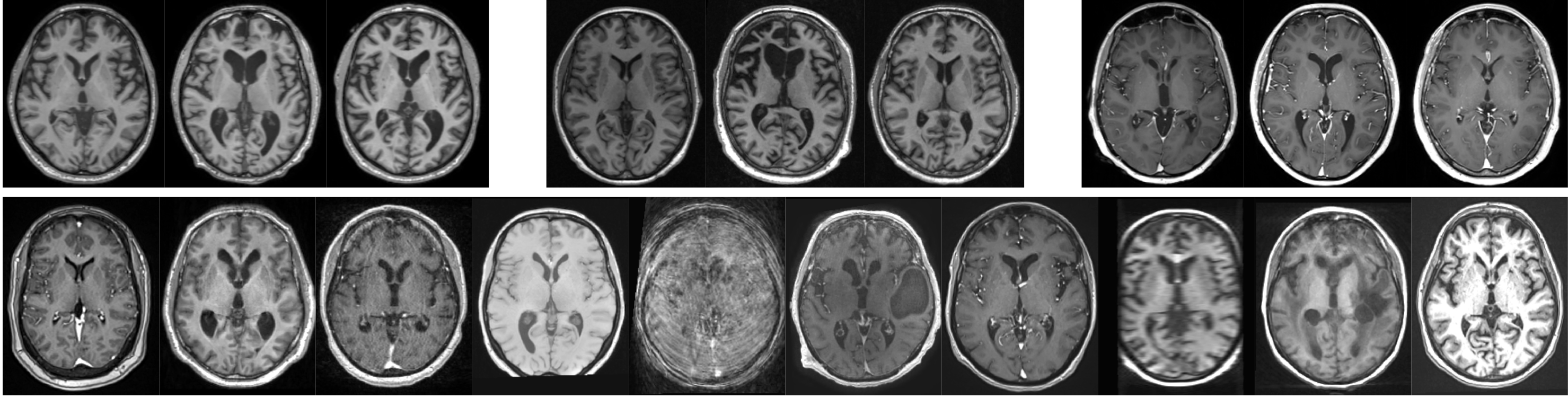
The PhD of Simona Bottani (2018–2022) laid the foundations of this line of work. Since exploiting clinical data warehouses for machine learning requires quality control (QC), and visual QC by experts is time-consuming and does not scale to large datasets, we developed a CNN for the automatic QC of 3D T1-weighted brain MRI from the AP-HP data warehouse, aiming to identify images that are not proper T1-weighted brain MRIs, detect gadolinium injection, and rate overall image quality (Bottani et al., Medical Image Analysis, 2022). Clinical data warehouses can also mix images acquired with and without a gadolinium-based contrast agent, which can bias results and limits the use of neuroimaging tools typically designed for non-contrast images only. We therefore explored image-to-image translation to convert contrast-enhanced into non-contrast-enhanced T1-weighted brain MRI, showing that reliable features could still be extracted from the synthetic images (Bottani et al., BMC Medical Imaging, 2024). Building on these quality-controlled and homogenised data, we then evaluated MRI-based machine learning approaches for computer-aided diagnosis of dementia on clinical routine data (Bottani et al., Medical Image Analysis, 2023). We showed that performance was strongly biased upwards by confounders of image quality and contrast agent injection, a phenomenon known as the Clever Hans effect or shortcut learning, and that once these biases were removed, performance remained considerably lower than on a research dataset.
This work was extended and consolidated during the PhD of Sophie Loizillon (2021–2024). A major contribution was the extension of our automatic quality control framework, first to improve the T1-weighted MRI tool using a transfer learning strategy based on synthetic artefact simulation (Loizillon et al., Medical Image Analysis, 2024; Loizillon et al., MELBA, 2024), and then to FLAIR MRI, a key sequence for the detection of white matter lesions widely used in clinical routine. We proposed a semi-supervised domain adaptation method that leverages the large pool of annotated T1-weighted images to minimise the annotation effort on FLAIR (Loizillon et al., Medical Image Analysis, 2025). A first step towards anomaly detection in clinical routine data was also taken by applying unsupervised anomaly detection using the β-VAE to FLAIR MRIs from the AP-HP data warehouse, targeting age-related white matter hyperintensities (Loizillon et al., MIDL, 2024). This feasibility study demonstrated that anomaly detection in routine care images is achievable, while also highlighting the additional challenges posed by data heterogeneity compared to images acquired in a research context.
Manon Heffernan started her PhD in 2024, pursuing this effort on FLAIR MRI quality control. She assessed whether the transfer learning strategy relying on artefact synthesis, which proved successful for T1-weighted MRI, could be transposed to FLAIR, and found that its performance was more limited in this setting (Heffernan et al., SPIE Medical Imaging, 2026). She also conductes a broader study of the quality of clinical routine T1-weighted and FLAIR brain MRI scans in the AP-HP data warehouse (Heffernan et al., OHBM, 2026).
Main publications
Quality control
- Bottani, S., Burgos, N., Maire, A., Wild, A., Ströer, S., Dormont, D., and Colliot, O.: Automatic Quality Control of Brain T1-Weighted Magnetic Resonance Images for a Clinical Data Warehouse. Medical Image Analysis, 75: 102219, 2022. doi:10.1016/j.media.2021.102219 HAL
- Loizillon, S., Bottani, S., Maire, A., Ströer, S., Dormont, D., Colliot, O., and Burgos, N.: Automatic Motion Artefact Detection in Brain T1-Weighted Magnetic Resonance Images from a Clinical Data Warehouse Using Synthetic Data. Medical Image Analysis, 93: 103073, 2024. doi:10.1016/j.media.2023.103073 HAL
- Loizillon, S., Bottani, S., Mabille, S., Jacob, Y., Maire, A., Ströer, S., Dormont, D., Colliot, O., and Burgos, N.: Automated MRI Quality Assessment of Brain T1-Weighted MRI in Clinical Data Warehouses: A Transfer Learning Approach Relying on Artefact Simulation. Machine Learning for Biomedical Imaging, 2(June 2024 issue): 888–915, 2024. doi:10.59275/j.melba.2024-7fgd HAL
- Loizillon, S., Bottani, S., Maire, A., Ströer, S., Chougar, L., Dormont, D., Colliot, O., and Burgos, N.: Automatic Quality Control of Brain 3D FLAIR MRIs for a Clinical Data Warehouse. Medical Image Analysis, 103: 103617, 2025. doi:10.1016/j.media.2025.103617 HAL
- Heffernan, M., Loizillon, S., Ströer, S., Chougar, L., Dormont, D., Colliot, O., and Burgos, N.: Assessing the Efficacy of Artefact Synthesis and Transfer Learning for Quality Control in Clinical 3D FLAIR Brain MRI. In Medical Imaging 2026: Clinical and Biomedical Imaging, 13929:57–64, SPIE, 2026. doi:10.1117/12.3085566 HAL
- Heffernan, M., Loizillon, S., Colliot, O., and Burgos, N.: Study of the Quality of Clinical Routine T1w and FLAIR Brain MRI Scans in a Clinical Data Warehouse. In OHBM 2026 – Annual Meeting of the Organization for Human Brain Mapping, Bordeaux, France, 2026. HAL
Dataset homogenisation
- Bottani, S., Thibeau-Sutre, E., Maire, A., Ströer, S., Dormont, D., Colliot, O., and Burgos, N.: Contrast-Enhanced to Non-Contrast-Enhanced Image Translation to Exploit a Clinical Data Warehouse of T1-Weighted Brain MRI. BMC Medical Imaging, 24(1): 67, 2024. doi:10.1186/s12880-024-01242-3 HAL
Computer-aided diagnosis of dementia
- Bottani, S., Burgos, N., Maire, A., Saracino, D., Ströer, S., Dormont, D., and Colliot, O.: Evaluation of MRI-Based Machine Learning Approaches for Computer-Aided Diagnosis of Dementia in a Clinical Data Warehouse. Medical Image Analysis, 89: 102903, 2023. doi:10.1016/j.media.2023.102903 HAL
- Loizillon, S., Jacob, Y., Maire, A., Dormont, D., Colliot, O., and Burgos, N.: Detecting Brain Anomalies in Clinical Routine with the β-VAE: Feasibility Study on Age-Related White Matter Hyperintensities. In Proceedings of the 7th International Conference on Medical Imaging with Deep Learning, 903–917, PMLR, 2024. PMLR HAL